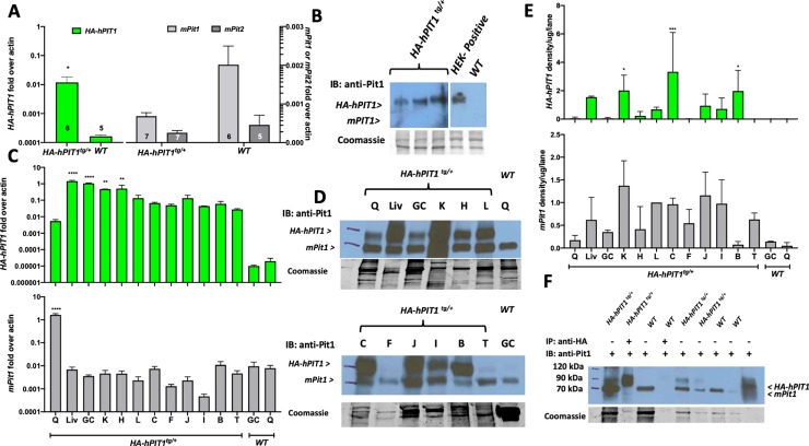
Fig 2. Expression of HA-hPIT1 and mPit1 in HA-hPIT1tg/+ and WT primary calvaria osteoblasts and various tissues.
(A) Semi-quantitative RT-PCR showing fold over actin expression in primary calvaria osteoblasts compared to wildtype at 80 days for hPIT1 and endogenous mouse Pit1 and 2 (mPit1 and 2). (B) Western blot of lysates obtained from PCOB from three separate HA-hPIT1tg/+ and one WT mouse using a lysate of HEK293 cells transfected with an expression plasmid encoding HA-hPIT1 as positive control, hybridized with anti-PIT1 rabbit polyclonal antibody (H-130, sc-98814, Santa Cruz Biotechnology) and detected with horseradish-peroxidase conjugated anti-rabbit IgG (mPit1 expected size 74 kDa, HA-hPIT1 expected size 75 kDa). (C) Semi-quantitative RT-PCR of total RNA from a HA-hPIT1tg/+ mouse shows expression of hPIT1 in different tissues (Liv = liver, K = kidney, C = colon, J = jejunum, I = ileum, B = brain, L = lung, T = testicle), compared to gastrocnemius (GC) and quadriceps (Q) from a wildtype (WT) mouse (n = 2) (D) Western blot of lysates from different tissues of a HA-hPIT1tg/+ and WT (hybridized with anti-PIT1 rabbit polyclonal antibody (H-130, sc-98814, Santa Cruz Biotechnology) and detected with horseradish-peroxidase conjugated anti-rabbit IgG) (mPit1 expected size 74 kDa, HA-hPIT1 expected size 75 kDa), (E) densitometric quantification of hPIT1 and mPit1 protein normalized to lung mPit1 and protein per lane from the Coomassie stains from two replicate Western blot experiments. (F) immunoprecipitation of HA-hPIT from quadriceps muscle obtained from a WT and HA-hPIT1tg/+ mouse. ****p<0.00002, ***p = 0.0002, **p = 0.002, *p = 0.03 vs. WT.

