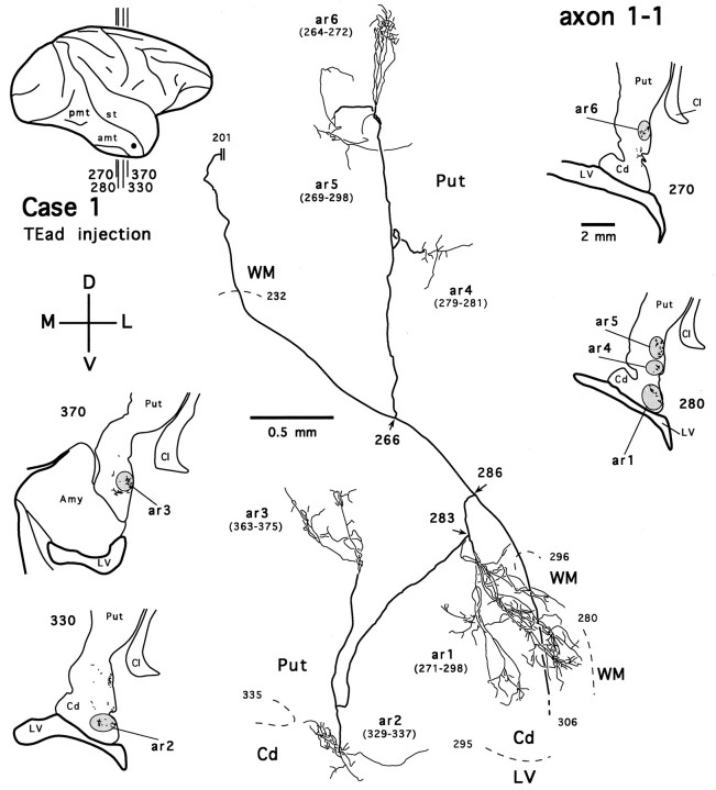
Fig. 14.
Camera-lucida reconstruction of axon 1-1 labeled by PHA-L anterogradely transported from TEad (case 1) to the ventrocaudal striatum. This axon was serially reconstructed through 112 sections (section thickness, 30 μm). Double linesindicate the incomplete portion of the axon. Abbreviations and conventions are the same as in Figure 7.

