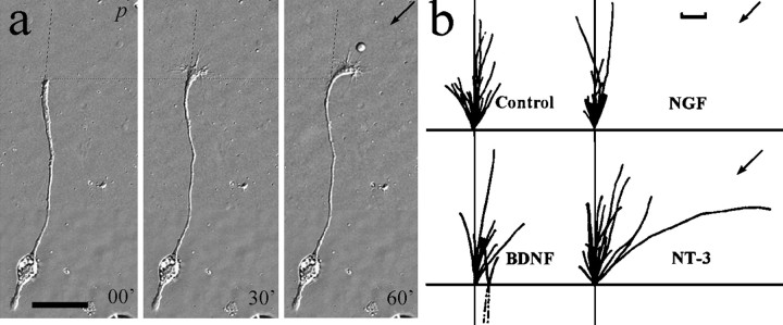
Fig. 8.
Turning response of 1-d-old Xenopusspinal neurons in the presence of neurotrophin gradients.a, Representative DIC images of a neuron at the onset and 30 and 60 min after the application of NT-3 gradient through a micropipette (p). Scale bar, 50 μm. Thedashed line indicates the original direction of neurite extension, and the dotted line indicates the corresponding positions along the neurite. b, Composite drawings of the path of neurite extension during a 1.5 hr period for all the neurons in the absence (control) and presence of different neurotrophins (50 μg/ml). The origin represents the position of the center of the growth cone palm at the beginning of the 1.5 hr experiment. The line depicts the trajectory of the neurite at the end of the 1.5 hr experiment. The arrowindicates the direction of the gradient. The initial direction of neurite extension (defined by the distal 20 μm segment of the neurite) was aligned with the vertical axis. In some cases the growth cone retracted slightly at the beginning of the experiment; for this reason some of the neurite drawings start withdashed lines. Scale bar, 10 μm.

