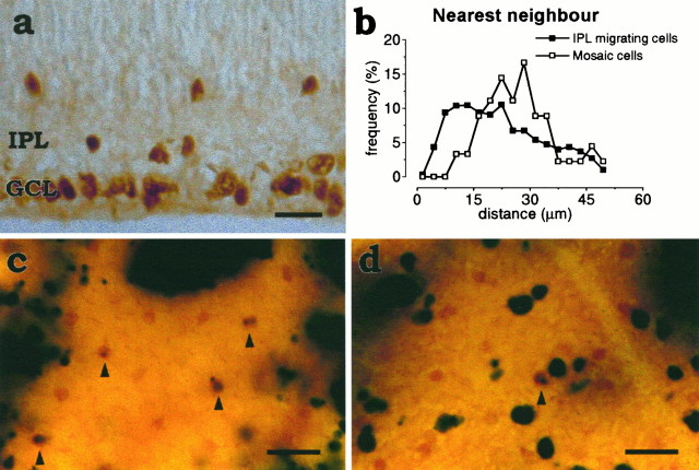
Fig. 3.
a, Islet-1+cells crossing the IPL have no minimal intercellular spacing. Cross section of a P1 retina. The spacing between Islet-1+amacrine cells migrating through the inner plexiform layer (IPL) was studied after P0 in the central retina, where the migration of ganglion cells that also express Islet-1 has ceased by this time. Scale bar, 10 μm. b, Lack of regular intercellular spacing between Islet-1 cells that are still outside the GCL mosaic. Normalized histogram of nearest neighbor distances between Islet-1+ cells in the IPL (filled squares) in P2 retinal sections. The histogram shows that the only limit to the spacing between Islet-1+ cells migrating through the IPL is the diameter of the cell, simply reflecting the fact that two cells cannot occupy the same physical space. The nearest neighbor distribution in the INL derived from the same sections (open circles) is shown as a comparison, because the minimal spacing is the same in both Islet-1 mosaics. As should be expected, the normalized distributions of nearest neighbor distances computed in sections and flat-mounted retina have the same minimal spacing but different tails, because the nearest neighbor in sections can be farther away (but, of course, never closer) than the real nearest neighbor in the mosaic. Counts were made in 30 randomly selected P2 sections from two retinae (n = 2).c, Retinal whole mount from an X-inactivation transgenic mouse showing a portion of the INL. Arrowheads point to four mosaic cells (brown; ChAT-immunoreactive) that are transgene-positive (blue) but are displaced in transgene-negative columns. These cells must be derived from neighboring transgene-positive clones. d, A portion of the ganglion cell layer showing one transgene-positive mosaic cell (arrowhead) and several transgene-positive ganglion cells (the largest cells in the field) in a transgene-negative column. These cells must have moved tangentially away from their clone of origin. Scale bar, 20 μm.

