Fig. 7.
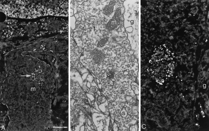
Transmission (B) and electron energy loss spectroscopic micrographs (A, C, ΔE = 150 ± 10 eV) of the cortical layer of the squid optic lobe (deep retina) showing carrot-shaped terminals of the optic nerve (bags). In A, several single and clustered ribosome-like signals (arrow) are seen in the bag outlined by adashed line. Magnification, 25,000×. Scale bar, 0.5 μm. The empty profiles within the bag are mitochondria (m). Identical ribosome-like signals are present in the rough endoplasmic reticulum of the nerve cell body located above the bag. A portion of the nucleus of the same neuron is shown at the top and is full of phosphorus signals derived from nucleosomal DNA. InC, a large aggregate of single and clustered ribosome-like signals is seen within a bag at a higher magnification (40,000×). By conventional EM (B), a carrot-shaped terminal of the optic nerve is characterized by the presence of several mitochondria, a host of synaptic vesicles, and small partially empty profiles representing the indentation of postsynaptic spines (s). Magnification, 25,000×. The arrow points to a long chain of dense spots resembling ribosomes. A glial process (g) is shown in B and C.

