
Fig. 3.
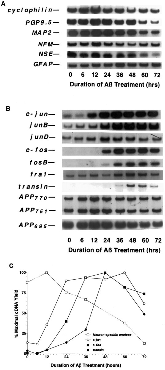
Time course of mRNA expression in rat cortical cultures undergoing Aβ-mediated neuronal apoptosis.A, Cellular marker genes. B, Jun and Fos family members and related genes. C, Quantification of changes in NSE, c-jun,c-fos, and transin expression. To assess changes in mRNA levels, we maintained primary rat cortical cultures (∼125,000 neurons/well) for 3–4 d and then treated them with Aβ1–40 (40 μm, lot ZM482), as described in Materials and Methods. After various times of Aβ treatment, total RNA was isolated, aliquots were converted to cDNA, and then 3% of the resultant cDNA was analyzed in each PCR sample. The data presented are from a single preparation of neuronal cultures, which were maintained and treated in parallel with those described in Figure 1. Each gene induction was confirmed in at least two independent neuronal culture preparations.