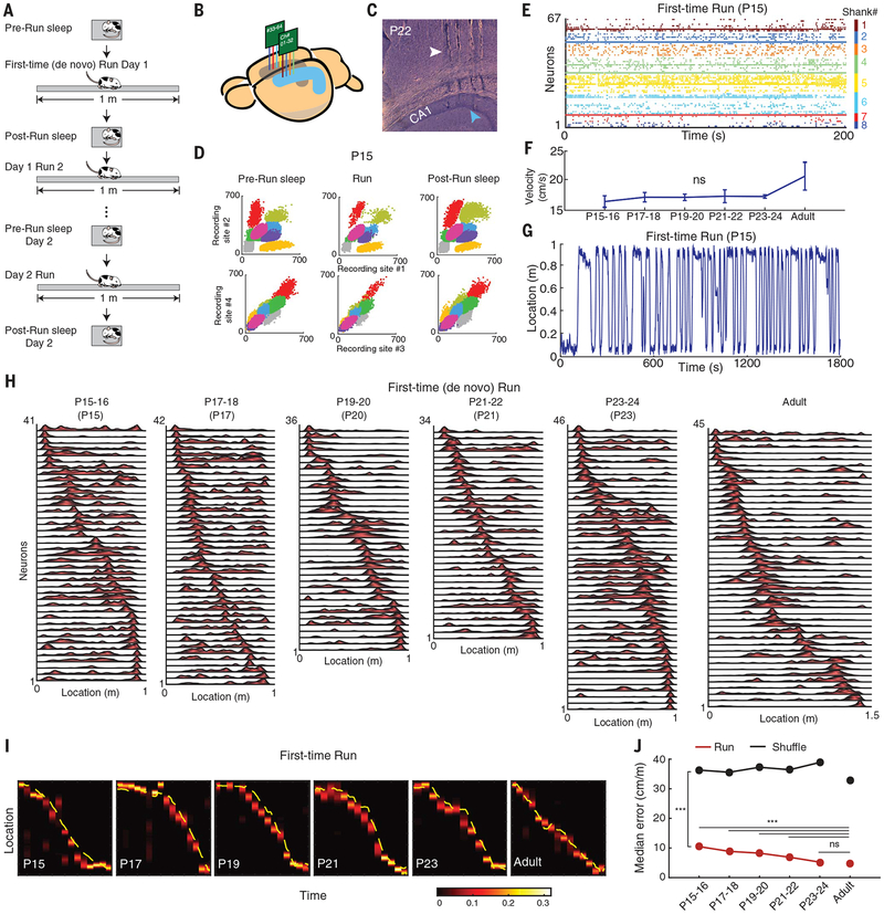
Fig. 1. Early-life development of hippocampal neuronal ensemble spatial representation during first-time navigation.
(A) Schematic of experimental protocol. (B) Illustration of bilateral silicon probe implants in the dorsal hippocampal area CA1. (C) Sagittal brain section depicting tracks of four silicon probe shanks (white arrowhead) recording in CA1 (blue arrowhead) at P22. (D) Color-coded illustration of clusters of eight single cells recorded from one shank in a P15 animal during sleep-run-sleep sessions. (E) Simultaneous recording of 67 CA1 neurons across eight shanks at P15. (F) Similar velocity during first-time (de novo) run across development [P = 0.209; analysis of variance (ANOVA)]. (G) Example of first-time run trajectory at P15. (H) Examples of simultaneously recorded place cell sequences during first-time run across development. (I) Increased accuracy in neuronal ensemble representation of spatial locations during first-time navigation across ages throughout development, based on a memoryless Bayesian decoding algorithm (heatmap). Yellow line: actual animal trajectory. The color map shows the probability of a decoded location. (J) Significance (from P15–P16 onward, run vs. shuffle; P < 10−10, rank sum tests) and age-dependent improvement (P < 10−10, Kruskal-Wallis ANOVA) of neuronal ensemble–decoded locations during first-time navigation across development. Representation becomes adult-like at P23–P24. For post hoc comparisons with adult data, Dunn’s test was used. ***P < 0.001; ns, not significant.

