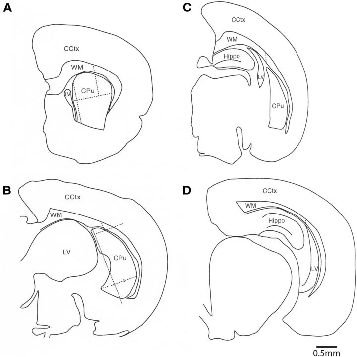
Figure 1.
Illustration of the boundaries used to define the callosal white matter. A–D are illustrations of coronal sections through a P4 rat brain proceeding from anterior (A) to posterior (D). The respective section numbers are 49 (A), 73 (B), 89 (C), and 113 (D), with all sections derived from the same rat. In all sections, the medial border of the callosal white matter (WM) was defined as the point at which the corpus callosum meets the edge of the section. In the sections anterior to the hippocampus (i.e., A, B), the lateral border of the callosal white matter was delineated by first measuring the maximum horizontal width of the striatum [i.e., caudate–putamen (CPu)] in each section. The vertical axis for this horizontal measurement was usually the vertical plane of the entire medial boundary of the caudate–putamen and its adjacent lateral ventricle (LV; see A). When the lateral ventricle abutted on less than one-third of the medial surface of the caudate–putamen, the superomedial boundary of the caudate–putamen was used as the vertical axis for this horizontal measurement (B). The horizontal measurement was completed on a carefully drawn camera lucida diagram of all the key brain regions in each section (A, B). Each section was carefully drawn by projecting it onto a whiteboard at 50×. The maximum horizontal width of the caudate–putamen was then divided by three to yield one-third of the maximum distance. This one-third distance was marked on the diagram from the most lateral edge of the caudate–putamen. A vertical line from this lateral one-third mark was then drawn superiorly through the white matter, in a plane that was parallel to the vertical line of the medial boundary of the caudate–putamen, to demarcate the lateral boundary of the callosal white matter (A, B). In the more posterior sections that contained the hippocampus, the lateral border was defined by first finding the most superior point of the caudate–putamen. A vertical line was then drawn superiorly from this point through the white matter, in a plane that was parallel to the vertical medial boundary of the caudate–putamen (C), to demarcate the lateral boundary of the callosal white matter. When the caudate–putamen was no longer present in the most posterior sections, the lateral border was defined by first finding the most superior point of the lateral ventricle. A vertical line was then drawn superiorly from this point through the white matter, in a plane that was parallel to the vertical medial boundary of the section (D). The white matter below the exclusion line was not sampled, whereas the white matter from above the line around to the medial edge of the corpus callosum was sampled. CCtx, Cerebral cortex; Hippo, hippocampus.

