. 2019 Oct 1;8:e47346. doi: 10.7554/eLife.47346

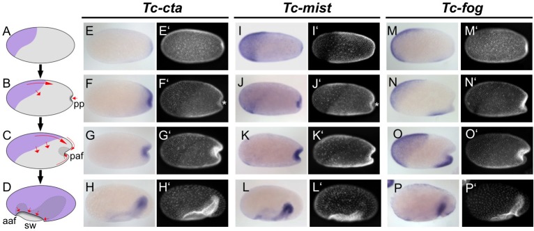
Figure 1. Expression of Fog signaling components during early embryogenesis.

(A–D) Schematics showing embryo condensation as described in the text. Serosa is shown in purple, germ rudiment tissue is shown in gray, arrows display tissue movements. aaf: anterior amniotic fold, paf: posterior amniotic fold, pp: primitive pit, sw: serosal window. (E–P’) Whole mount ISH and DNA staining for Tc-cta (E–H), Tc-mist (I–L) and Tc-fog (M–P). (E’–P’) nuclear (DAPI) staining of respective embryos. Anterior is left, ventral is down (where possible to discern).

Figure 1.

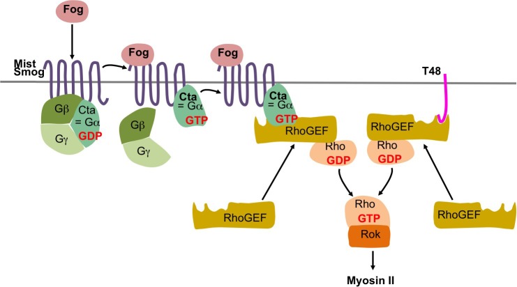
Figure 1—figure supplement 1. Fog and T48 pathway in Drosophila.

Figure 1—figure supplement 1.

In Drosophila the secreted protein Fog activates the two G protein coupled receptors (GPCRs) Mist (Mesoderm-invagination signal transducer, also known as Mthl1 (Methuselah-like1)) (Manning et al., 2013) and Smog (Jha et al., 2018; Kerridge et al., 2016). The active receptors act through a heterotrimeric G protein composed of Gα12/13, Gβ13F, and Gγ1 (Kerridge et al., 2016) by converting the Gα12/13 subunit, known as Concertina (Cta) from its GDP bound inactive to its GTP bound active form. The complex then dissociates into free Gα12/13 and a Gβ13F/Gγ1 dimer, which regulate their respective intracellular effectors. In particular, active Gα12/13 recruits RhoGEF2 to the apical plasma membrane where it acts as a guanine nucleotide exchanges factor for Rho1. Rho1-GTP then activates Rok (Rho kinase). Rok phosphorylates the regulatory light chain of non-muscle myosin II promoting apical myosin contractility. RhoGEF2 can also be recruited to the apical membrane through the transmembrane protein T48 which interacts with RhoGEF2 via its PDZ-binding domain (Kölsch et al., 2007).
Figure 1—figure supplement 2. Insect Fog proteins.
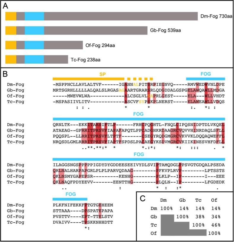
Figure 1—figure supplement 2.

(A) Schematic representation of Fog proteins from Drosophila melanogaster (Dm-Fog), Gryllus bimaculatus (Gb-Fog), Oncopeltus fasciatus (Of-Fog) and Tribolium castaneum (Tc-Fog). The proteins contain an N-terminal signal peptide (yellow) followed by a conserved Fog domain (blue) and a highly variable C-terminal domain. (B) Sequences of the N-terminal regions including the signal peptide (SP, yellow) and the Fog domain (FOG, blue). Amino acids marked in yellow indicate the predicted cleavage side of the SP (identified via SignalP 4.1 (http://www.cbs.dtu.dk/services/SignalP/)). (C) Sequence conservation within the Fog domain as indicated as percentage of identical amino acids.
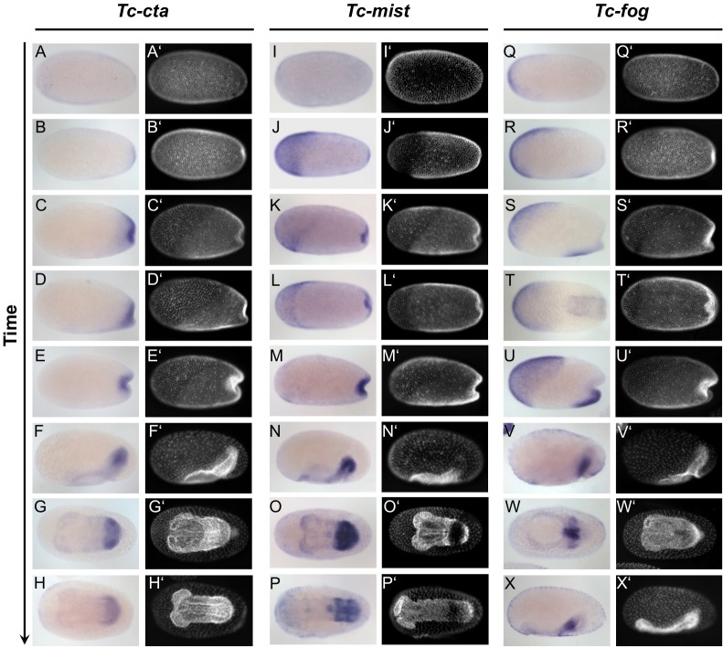
Figure 1—figure supplement 3. Expression of Tc-cta, Tc-mist and Tc-fog during early embryogenesis.

Figure 1—figure supplement 3.

(A–X) Whole mount ISH for Tc-cta (A–H), Tc-mist (I–P) and Tc-fog (Q–X). (A’–X’) DAPI staining of respective embryos. Anterior is left, all panels show optical sagittal sections, except G, H, L, O, P, T and W, which show ventral surface views.
Figure 1—figure supplement 4. Tc-fog and Tc-twi are co-expressed only within the posterior presumptive mesoderm.
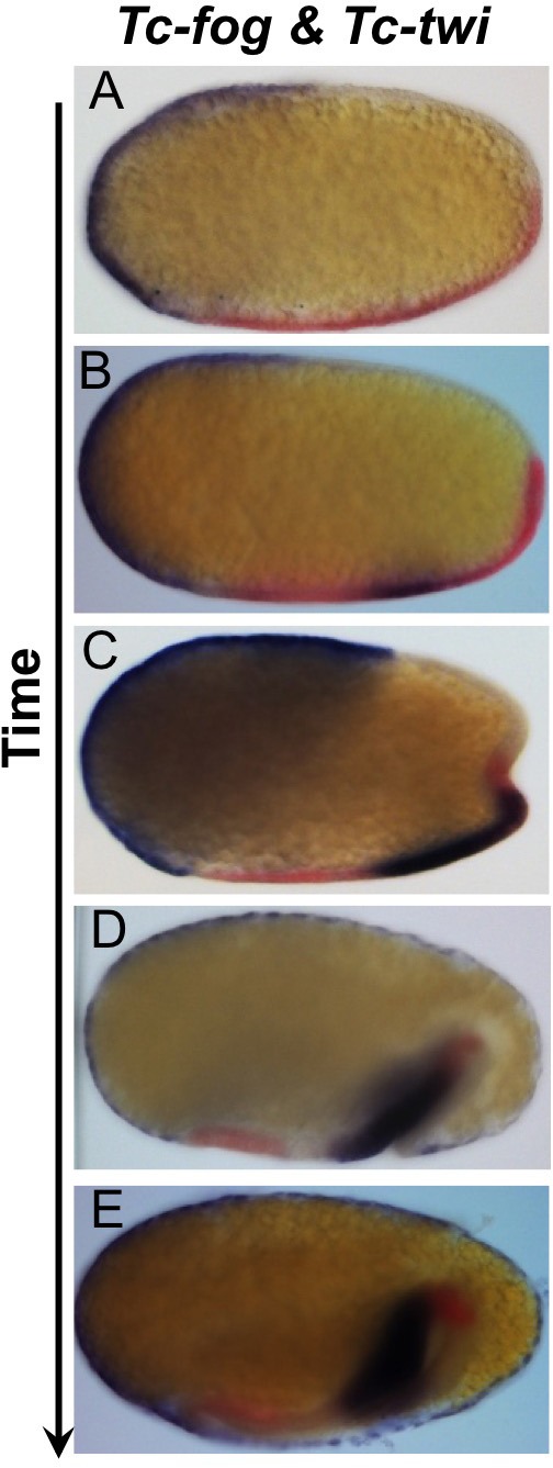
Figure 1—figure supplement 4.

Double whole mount ISH for Tc-twi (red) and Tc-fog (blue). Anterior is left, all panels show optical sagittal sections.