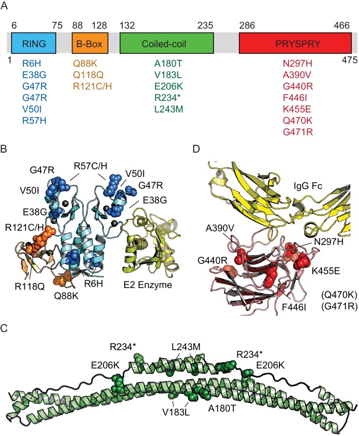
Figure 2. Domain location of TRIM21 missense variants.
(A) Schematic of TRIM21 showing how variants are distributed amongst component domains. (B–D) Mapping of variants onto structures of each domain. (B) RING (blue) and B-Box (orange) variants are marked on the autoinhibited structure of the TRIM21 RING-B-Box domains (PDB 5OLM). The second copy of the B-Box has been omitted and the location of a bound E2 enzyme (yellow) has been included instead to indicate the location of this functional interface (based on superposition of TRIM25 RING: E2 structure (5FER)). (C) Coiled-coil domain (green) based on TRIM25 (PDB 4CFG). Location of TRIM21 residues are marked based on sequence alignment. (D) PRYSPRY domain (red) with bound IgG Fc (yellow) based on PDB 2IWG. Residues in brackets are not present in the structure.

