. 2019 Oct 15;8:e50069. doi: 10.7554/eLife.50069

Figure 4. Evidence for ROS-related oxidative protein damage in GRU102.

(A) Densitometric analysis of protein carbonyl content (PCC) from whole lysate in young GRU101 and GRU102. (B) Densitometric analysis of mitochondrial protein carbonyl content (mtPCC) from whole lysate in young GRU101 and GRU102 (unpaired t-test, p<0.05, *; n = 3–7 repeats per condition, with approximately 1500 animals per repeat collected from independent cohorts).

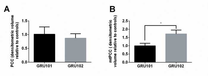
Figure 4—source data 1. Protein carbonyl content (PCC) in D4 GRU101 and GRU102.
All values were normalized to D4 GRU101.
DOI: 10.7554/eLife.50069.014

Figure 4.

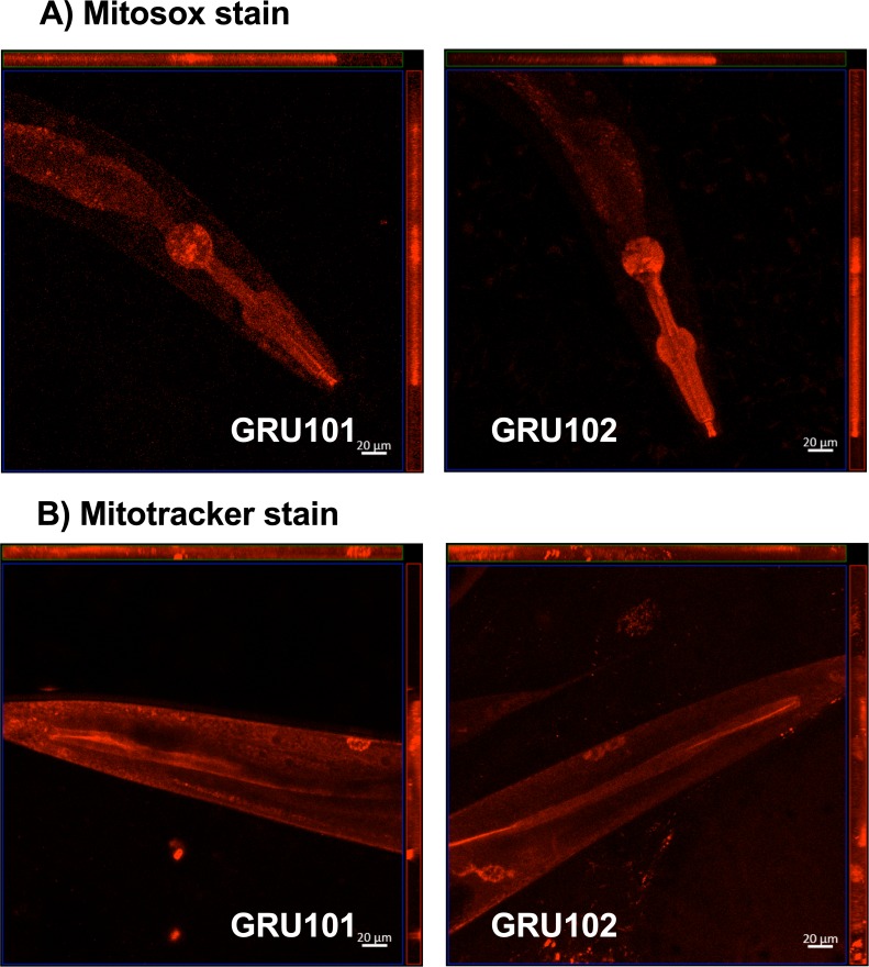
Figure 4—figure supplement 1. Mitosox and mitotracker staining of GRU101 and GRU102 animals.

Figure 4—figure supplement 1.

(A) Mitosox stain was performed on day-4 animals, when reactive oxygen species (ROS)-mediated protein damage was observed. (B) Mitotracker stain was performed on day-10 animals, when impairment in mitochondrial health (reduction in oxygen consumption capacities measured by Seahorse metabolic flux) was first observed.