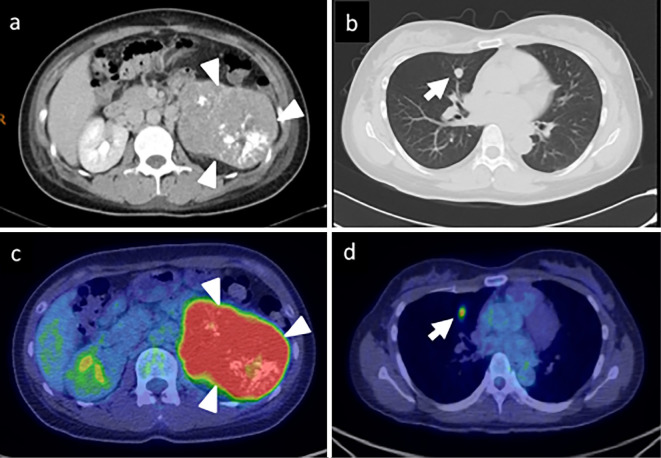
Figure 1.
a, b) Contrast-enhanced computed tomography (CT) showing a large left adrenal tumor with heterogeneous enhancement (arrowhead) and a right pulmonary tumor (arrow). c, d) Positron 18F-fluorodeoxyglucose positron emission tomography with CT showing both tumors, with maximum standardized uptake values of 9.5 and 3.9.

