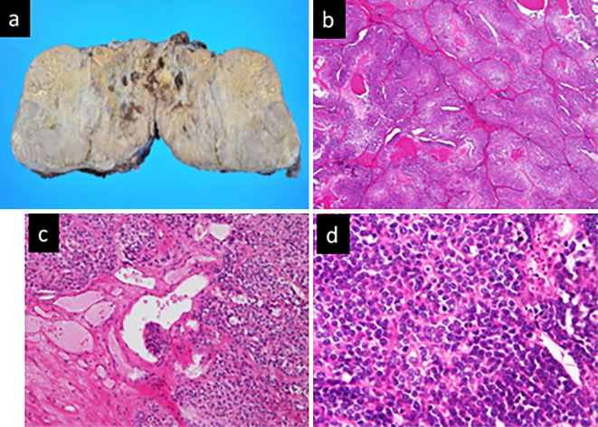
Figure 2.
Adrenocortical carcinoma of the left adrenal gland. a: Resected specimen. b: Clusters of tumor cells with eosinophilic cytoplasm and <25% clear cells [Hematoxylin and Eosin (H&E) staining, ×40]. c: Sinusoidal invasion (H&E staining, ×200). d: Mitotic rate >5/50 high-power fields (H&E staining, ×400).

