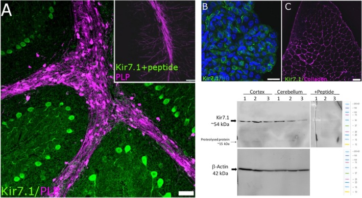
Figure 1.

Validation of Kir7.1 immunostaining. (A‐C) To serve as positive controls, immunostaining for Kir7.1 is demonstrated in Purkinje neurones (A; adult PLP‐DsRed mouse cerebellum, in which white matter tracts appear magenta) and choroid plexus epithelium (B; counterstained with Hoechst Blue to visualise the cell nuclei). Immunostaining was absent following pre‐incubation in blocking peptide (A, Inset), and in adult mouse skeletal muscle, which does not express Kir7.1 (C; counterstained for collagen, which appears magenta). (D) Western blot analysis of protein lysates from mouse cerebellum (CBL) and cortex (CTX) confirmed robust Kir7.1 protein expression with a predicted band at 54 kDa. Positive bands were absent in the presence of the competitive peptide. In some samples, very dim bands were observed at approximately 15 kDa, which corresponds to protein proteolysed during sampling. Scale bars: (A,C) 50 μm; (B) 10 μm; (A, Inset) 100 μm.
