
Figure 3.
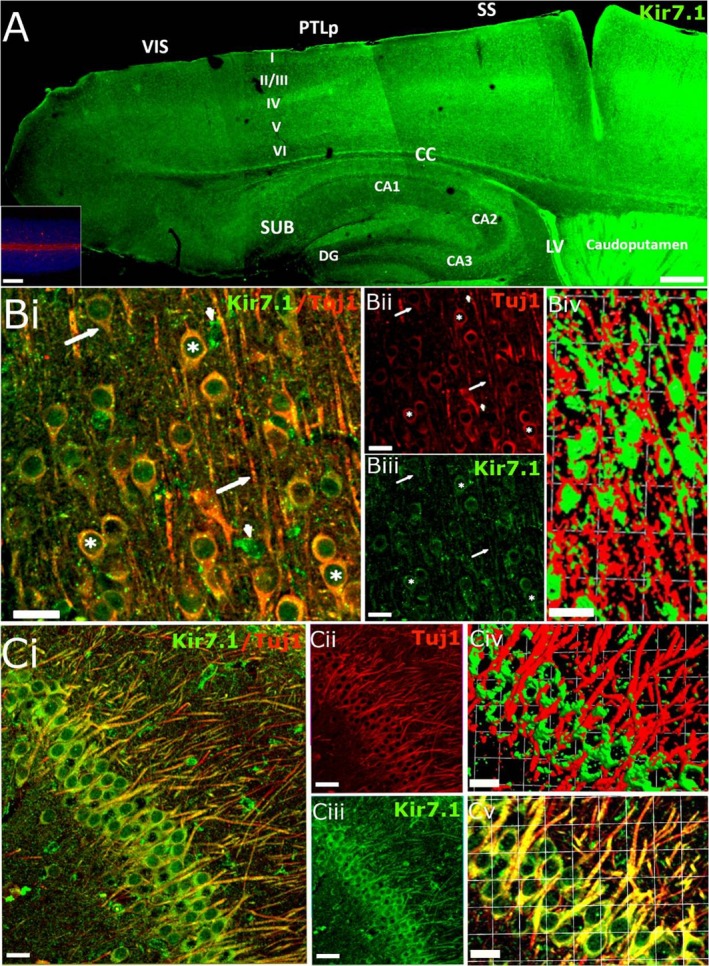
Kir7.1 immunostaining in the adult mouse forebrain. (A) Overview of the pattern of Kir7.1 expression in the adult mouse forebrain. Kir7.1 immunostaining was absent in negative controls pre‐incubated with peptide (A, Inset). (B,C) Double immunofluorescence labelling for Kir7.1 (green) and Tuj1 (red) in the cortex (B) and hippocampus (C). (B) Layer 2/3 cortical neurones expressing Kir7.1 on their cell bodies (asterisks) and axons (arrows); co‐expression appears yellow in the overlay (Bi) and Kir7.1 immunopositivity can be seen in Tuj1‐negative cells, which are likely to be astrocytes (arrowheads); individual channels are illustrated for Kir7.1 (Bii) and Tuj1 (Biii), together with isosurface images showing the close apposition of Kir7.1 and Tuj1 voxels (Biv). (C) Hippocampal pyramidal cells express Kir7.1 on cell somata and axons (Ci); individual channels are illustrated for Kir7.1 (Cii) and Tuj1 (Ciii), together with higher magnification of the overlay image (Cv) and isosurface image (Civ), illustrating the close apposition of Kir7.1 and Tuj1 voxels. Scale bars: (A) 300 μm; (A, Inset) 100 μm; (Bi‐iii and Ci‐iii) 25 μm; (Biv) 1 square unit = 35.42 μm; (Civ‐v) 1 square unit = 16.64 μm.