
Figure 6.
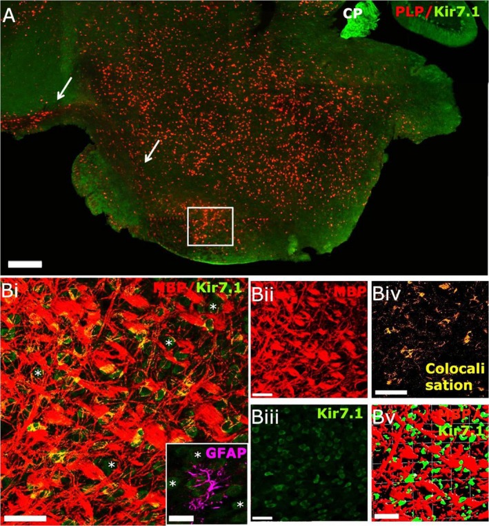
Kir7.1 expression in the mouse hindbrain. (A) Low‐magnification confocal image of sagittal section from adult PLP‐DsRed (red) reporter mouse immunolabelled for Kir7.1 (green). Kir7.1 is widely expressed in the pons, with intense expression in the choroid plexus of the third ventricle (CP), whereas the corticospinal tract appears immunonegative for Kir7.1 (arrows). (B) Higher magnification images from the area indicated by the white square in (A), immunostained for MBP (red) and Kir7.1 (green), showing neuronal expression of Kir7.1 (asterisks) and some co‐localisation of Kir7.1 and MBP(appears yellow in Bi); individual channels are illustrated for MBP (Bii) and Kir7.1 (Biii), together with the colocalisation channel (Biv) and isosurfacing image (Bv), which does not reveal very close apposition of Kir7.1 and MBP voxels (Bv), suggesting that Kir7.1 may be expressed by axons and not by myelin. (Bi‐inset) Double immunolabelling for Kir7.1 (green) and GFAP (magenta) did not detect expression of Kir7.1 in astrocytes of the pons. Scale bars: (A) 300 μm; (Bi‐v) 50 μm; (Bi, Inset) 25 μm.