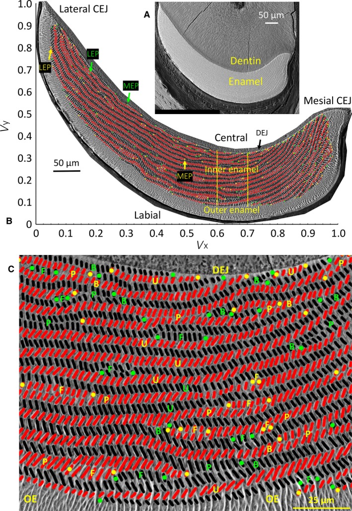
Figure 1.

Backscattered scanning electron microscopic (BEI) images of a right mandibular mouse incisor in transverse section viewed looking in an apical direction (toward growing apical end) and photographed at low and high magnification (A, B), and a cropped portion of the inner enamel layer from the central labial side of the incisor (C). (A) Single low‐magnification BEI image of labial side of a mandibular mouse incisor in a typical transverse section of the tooth showing the location of dentin and enamel. The cracks in the dentin are an artifact caused by air drying the tissue slice after polishing. (B) A high‐resolution map of the same tooth section shown in (A) made from BEI images photographed at × 800 and montaged to recreate the whole enamel layer. The cut open oval profiles of enamel rods are arranged in rows across the inner enamel layer between the limits of the mesial and lateral cementoenamel junctions (CEJ). The enamel rods are colored black for rows having a mesial tilt and red for rows having a lateral tilt. The mesial endpoints (MEP) and lateral endpoints (LEP) of the rows are color‐coded green for rows having a mesial tilt and yellow for rows having a lateral tilt. Superimposed over the image are the x‐ and y‐axes of a graph illustrating the normalized virtual coordinate system (V x, V y) used for defining the locations of individual rods or regions of the inner enamel layer: lateral region to the left of the vertical yellow line, central region within the two vertical yellow lines, and mesial region to the right of the second vertical yellow line (see Table 1 for classification details). (C) The rows of enamel rods show a variety of arrangements, including uniform appearance along their length with opposite tilt angles in rows above and below (u), sites where two rows with the same tilt come into close proximity to each other (paired) (p), rows that appear to branch into two rows with the same tilt or where two rows with the same tilt merge into one row (B), and sites where petite rows appear located at the sides of longer rows having the same tilt (called focal stacks, F). DEJ, dentinoenamel junction; OE, outer enamel.
