Figure 7.

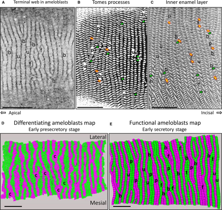
One‐micrometer‐thick tangential sections (plastic) of secretory stage rat incisor ameloblasts at the level of their distal terminal webs (A) and at the level of the Tomes processes projecting into the forming enamel layer (B), and in nearly mature enamel from the maturation stage (C) stained with toluidine blue. The tilt of a row cannot be defined in (A), but uniform rows (u), branching/merging rows (B) and sites where a focal stack might be located (F) are apparent. Tomes processes projecting into the forming enamel layer from secretory stage ameloblasts (B) show many of the row arrangements typically identified later in enamel rod arrangements (C) that develop from these distal extensions of the ameloblasts. This includes uniform rows (u), branching/merging rows (b), paired rows (p) and focal stacks (f). In (B) and (C), the endpoint for rows having a mesial tilt is indicated in green with black outline, and for rows having a lateral tilt in yellow with red outline. The colored maps in (D) and (E) are graphic reconstructions from serial sections (see Materials and methods) of the row arrangement of ameloblasts at the level of the distal terminal web in young differentiating ameloblasts (D) and in early secretory stage ameloblasts (E). The development of rows of ameloblasts clearly begins very early as pre‐ameloblasts start to differentiate (d) with focal areas where cells seem clustered (c) at sites that may have something to do with branching/merging of rows seen more clearly later in time (D). As secretion of the enamel begins (E), row structure is more regular and spread out as uniform (u), paired (p), branching/merging (b) and focal stack (f) patterns. In all panels the magnification bars = 25 μm, apical is to the left, incisal to the right, lateral to the top, and mesial to the bottom.
