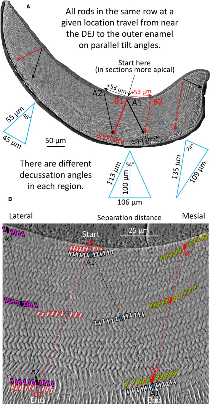
Figure 8.

Schematic illustrations of enamel rod decussation taking into account increases in the mean decussation angle from lateral to mesial regions of the inner enamel layer (46°, 54°, 74°; Smith et al. 2019) for the whole enamel layer (A) and central region only (B). Rows of ameloblasts that form the rows of enamel rods undergo their differentiation as the wave of tooth development spreads from the central region toward the mesial and lateral regions of the future enamel layer (e.g. red and black arrows labeled + 53 μm in top figure). Rows therefore are created and lengthen at sites close to the DEJ. A transverse section of the mature enamel layer is a time composite image of all the various generations of ameloblasts that were required to create the rows of rods layered one‐on‐top another. This implies that two neighboring ameloblasts, one from a row destined to form an enamel rod having a mesial tilt (black, A1) and the other from a row creating enamel rods having a lateral tilt (red, B1) spread apart by a distance that can be estimated from planar geometry using decussation angle and the thickness of the enamel layer over which the cells will travel (blue triangles). As these ameloblasts and their sister cells in the same row move to form a corresponding row of enamel rods, new ameloblasts continue to develop near the DEJ along the same row or as part of new rows having similar rod tilts (A2 and B2). At the boundary area where ameloblasts switch from forming the decussating inner enamel portions of the rods to forming the parallel outer enamel portions of the rods, ameloblast A1 will oppose ameloblast B2 that differentiated at a more mesial spatial location than ameloblast B1, and ameloblast B1 will oppose ameloblast A2 that differentiated at a more lateral location than ameloblast A1. This implies that the potential space that would arise as ameloblasts in row A physically move in a mesial direction over space and time from ameloblasts in row B moving laterally is pre‐compensated for by transverse extensions of arcing rows and creation of new rows as the wave of amelogenesis spreads into the mesial and lateral regions along the DEJ. It is important to keep in mind that because enamel rods are tilted incisally (forwards) at about 45° to the DEJ in the plane of tooth eruption, the START positions for rows of rods at the DEJ in 3D space are positioned at least 100 μm more apically (backwards into the plane of the photographs shown here) than their END locations at the boundary with the outer enamel layer. It is not possible to see the start and end positions of an enamel rod in a single 2D section as represented in this figure, which is strictly for conceptual purposes. DEJ, dentinoenamel junction.
