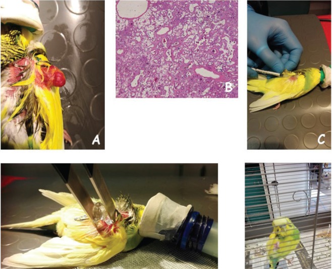
Fig. 1. (A) The patient at presentation showing a large, ulcerate, exophytic mass on the left wing. (B) The histological appearance of the tumor made up by a mixture of areas of epithelioid hemangioendothelioma and areas of low-grade angiosarcoma (H&E; original magnification × 10). (C) Bleomycin is locally injected in the tumor bed. (D) Permeabilizing electric pulses are administered using plate electrodes. (E) The patient at 3-months follow-up showing no evidence of disease.

