Figure 7.
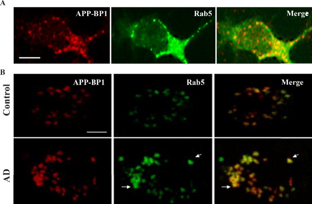
APP–BP1 and Rab5 colocalize in cultured neurons overexpressing APP–BP1 and in neurons in control and AD frontal cortex, as shown by confocal microscopy. A, Immunocytochemical analysis of neuronal cultures expressing exogenous APP–BP1 revealed close to a 63% overlap in their subcellular distributions, primarily in punctuate endosome-like structures. Scale bar, 5 μm. B, To determine whether these data are reflected in vivo, we stained postmortem prefrontal cortex tissue from AD and control human brains with antibodies to APP–BP1 and Rab5. The staining reveals an average of 82% overlap between APP–BP1 and Rab5 in control and AD brain, particularly in the enlarged endosomes in AD (arrows). Scale bar, 5 μm. Overlap was determined by comparing the number of costained cells with the total number of stained cells.

