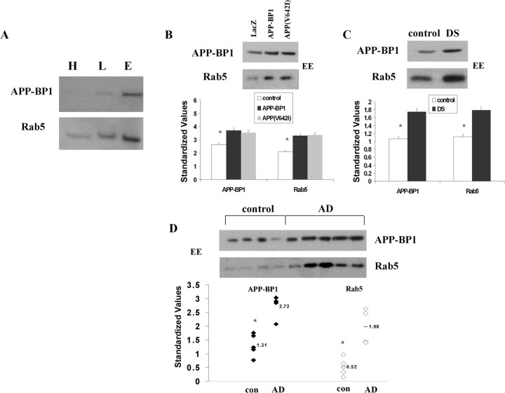
Figure 8.
APP–BP1 and Rab5 levels are increased relative to controls in early endosomes in primary neurons expressing exogenous APP–BP1 or APP(V642I), in DS skin fibroblasts, and in AD hippocampus. A, Subcellular endosomal fractionation of rat brain tissue shows increased levels of APP–BP1 specifically in the Rab5-positive early endosome fraction. H, Heavy membranes; L, late endosomes; E, early endosomes. B, Primary cortical rat embryonic neurons expressing APP–BP1 or APP(V642I) were fractionated and subjected to immunoblot analysis. Equal amounts of protein were loaded for all samples, and densitometry values were standardized to levels of GAPDH. Neurons expressing APP–BP1 or APP(V642I) show an increase in standardized APP–BP1 and Rab5 levels in early endosomes (EE) relative to controls (neurons expressing LacZ). C, D, Lysates of skin fibroblasts from DS subjects (C) and from postmortem hippocampal tissue from control and AD brains (D) were similarly fractionated and subjected to immunoblot analysis. In D, each lane represents a postmortem tissue sample from a distinct control (con) or AD subject (see Table 1). ANOVA showed significant increases in standardized levels of APP–BP1 and Rab5 in early endosomes in DS cells and in AD brain relative to controls. *p < 0.001.

