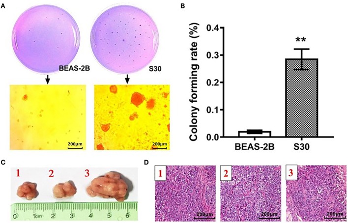
Figure 1.
CS-induced malignant transformation in BEAS-2B cells in vitro and in vivo. (A) Representative photographs of the colony formation assay of the normal BEAS-2B cells and S30 cells. (B) Graph of soft agar colony forming rate of normal BEAS-2B cells and S30 cells. **p < 0.01 vs. BEAS-2B. (C) Photographs of tumors excised 45 days after injection of normal BEAS-2B cells and S30 cells into nude mice. (D) Representative HE staining histopathologic image of tumor tissues excised 45 days after injection of S30 cells into nude mice.

