
Figure 4.
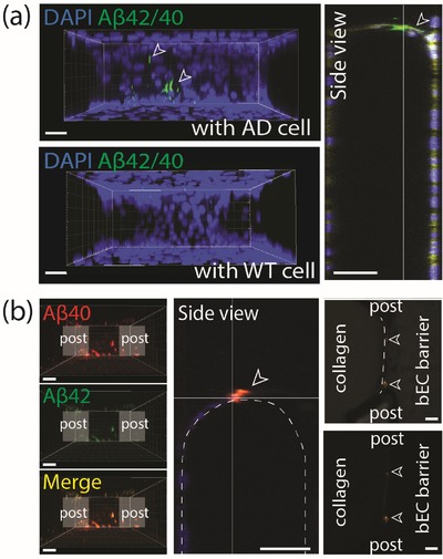
Aβ deposition in the AD model. a) Deposition of Aβ (green) on the BBB monolayer determined by immunofluorescence staining with Aβ 42/40 antibody. White arrows indicate deposited Aβ on the bEC barrier. Scale bars: 30 µm. b) Deposition of flour 555‐labeled Aβ 40 and FAM‐labeled Aβ 42 which were introduced into ReN cell media MC, on the bEC barrier. White arrows indicate deposited Aβ on the bEC barrier. Scale bars: 50 µm.