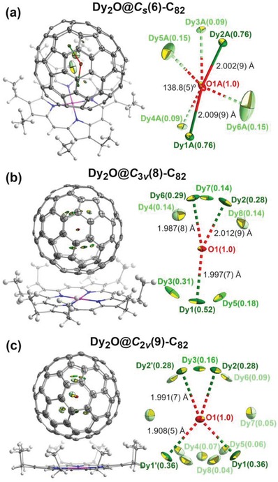
Figure 2.

Single‐crystal X‐ray structure of Dy2O@C82 isomers co‐crystallized with Ni(OEP): a) Dy2O@C s(6)‐C82; b) Dy2O@C 3v(8)‐C82; c) Dy2O@C 2v(9)‐C82. For each isomer, the structure of Dy2O@C82 ∙ Ni(OEP) is shown on the left (structures are oriented so that molecular symmetry plane is parallel to the paper), and enlargement of the endohedral Dy2O unit with disordered Dy sites and selected structural parameters is shown on the right. The brightness of the color differentiates the site occupancies (the darker the color, the higher the occupancy). Solvent molecules are omitted for clarity. The displacement parameters are shown at the 10% probability level. Color code: grey for carbon, green for Dy, red for O, blue for N, white for H, and purple for Ni.
