Fig. 1.
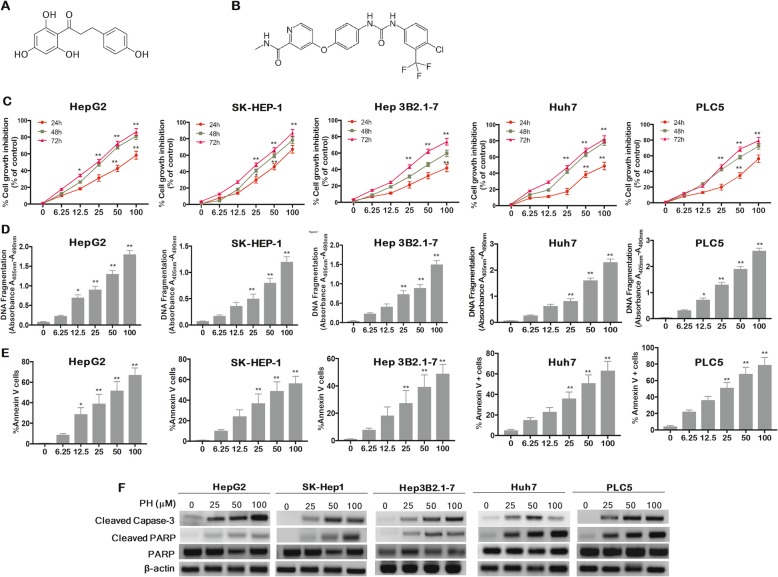
PH inhibits cell proliferation and induces apoptosis in HCCs. a Sor and (b) PH structure. c Cell viability by MTT assay. d DNA fragmentation assay. e Apoptosis was analyzed by annexin V, FACS analysis. Annexin V (+) cells were quantified. f The protein levels of caspase-3 and PARP were determined by Western blot after exposing HCCs to PH for 48 h. Experiments were conducted in triplicate and mean values ± SD (bars) are shown. *p < 0.05, and **p < 0.01 compared to control

