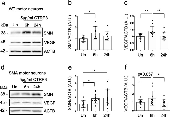
Fig. 4.
CTRP3 increases SMN and VEGF protein levels in primary WT and SMA motor neurons. Representative Western blots and quantification of SMN and VEGF protein levels in CTRP3 treated motor neurons isolated from spinal cord of E13 mice. Embryonic motor neurons from WT (a-c) and SMA (d-f) mice were cultured for 5 days in vitro (5DIV) and treated with 5 μg/ml mCTRP3 protein for 6 or 24 h. b, c, e and f Dot plot bar graphs represent quantification of VEGF and SMN levels. ACTB was used as a loading control. Each dot represents independent experiment and the bar graph represents mean ± s.d. (WT-SMN: n = 11, N = 5; WT-VEGF: n = 7, N = 3; SMA-SMN: n = 8; N = 3; SMA-VEGF: n = 11, N = 5, n: number of independent experiments, N: number of independent neuron cultures) One way ANOVA with Fisher’s LSD test was used to determine statistical significance; * p < 0.05, ** p < 0.01

