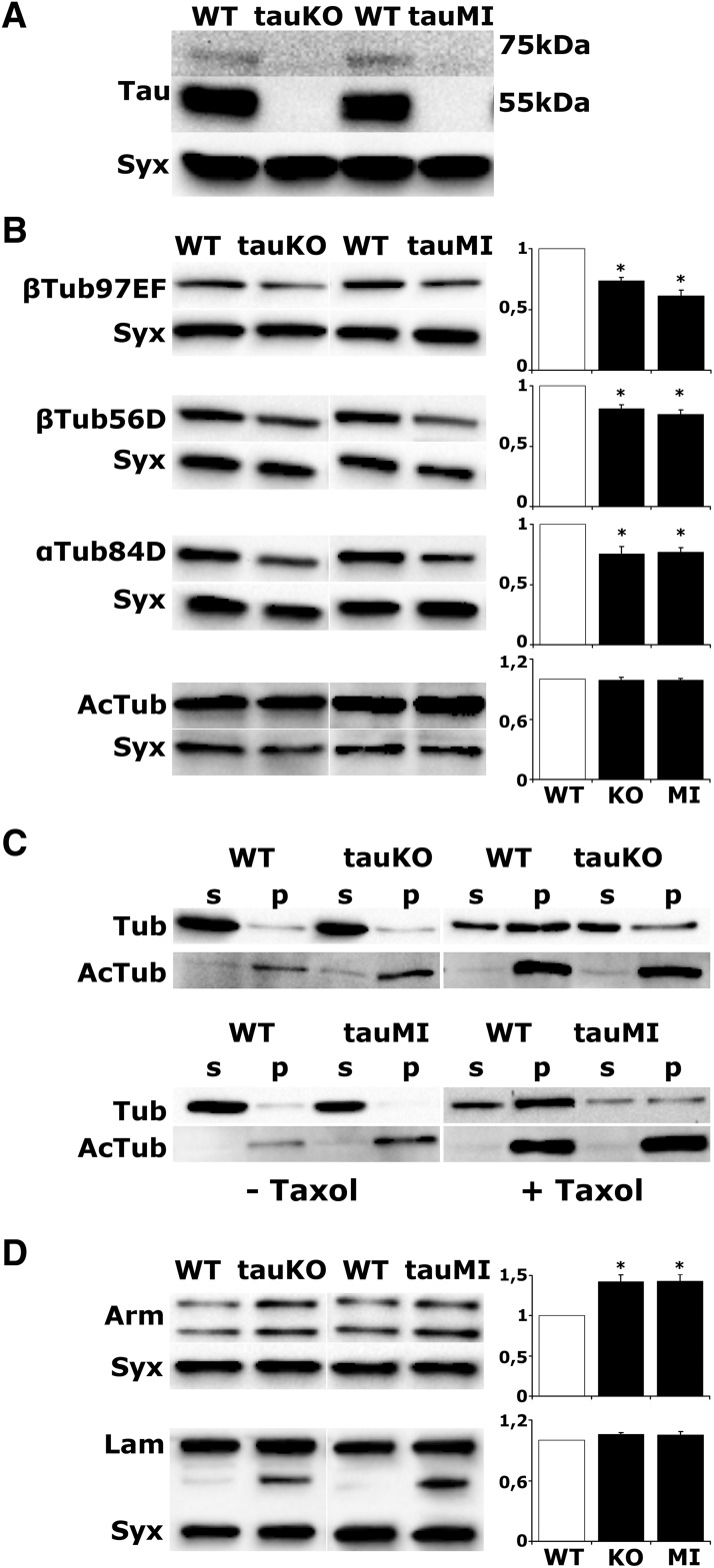
Figure 1.
dTau loss precipitates changes in microtubule cytoskeleton. A, Western blot analysis of head lysates from WT, tauKO, and tauMI flies probed with anti-dTau. B, D, Representative blots of head lysates probed with the indicated antibodies. For quantifications, levels of the indicated protein in the mutants were normalized using the syntaxin (Syx) loading control and are shown as a ratio of their mean ± SEM values relative to their respective level in WT flies, which is arbitrarily set to 1. Stars indicate significant differences (p < 0.05) from control (open bars) for tauKO and tauMI. C, Endogenous microtubules were purified from fly head lysates in the absence or presence of Taxol. p, Pellet fraction; s, supernatant fraction. Fractions were analyzed by Western blotting using antibodies against total tubulin (Tub) and acetylated tubulin (AcTub).

