Figure 1.

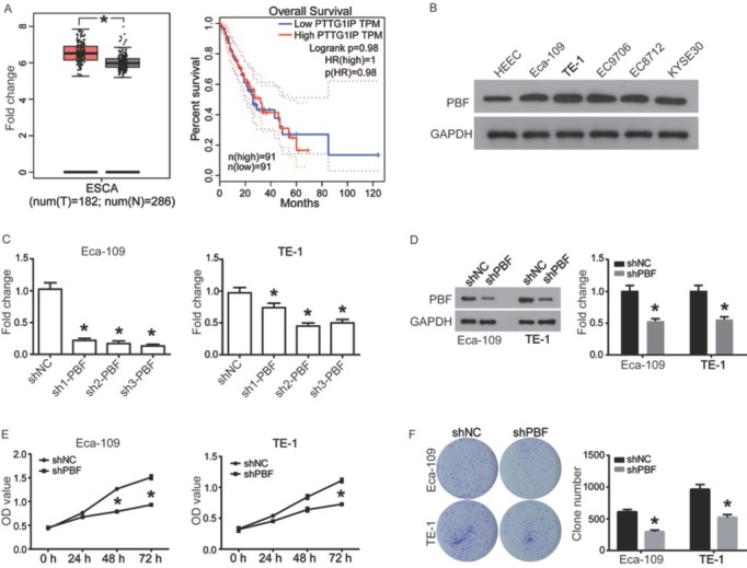
PBF was highly expressed in ESCA and down-regulation of PBF significantly inhibits proliferation of ESCAS cells. (A) Left: The expression of PBF in ESCA tissue (n=182) compared with normal controls (n=286); The red and gray boxes represent ESCA tissue and normal tissues, respectively; Right: The correlation of PBF mRNA expression with the overall survival of patients with ESCA; (B) PBF protein expression was investigated in human normal esophageal epithelial cell line HEEC and ESCA cell lines Eca109, TE-1, EC9706, EC8712 and KYSE30 cells by western blot analysis. (C)RT-qPCR was utilized for detecting mRNA expression of PBF in ESCA cells transfected with shNC or shPBFs for 24 h; (D) Western blot was used to confirm the interference effect of sh2-PBF on PBF protein level. (E) CCK8 assay was utilized for analyzing cell proliferation of Eca-109 and TE-1 cells, which were pre-transfected with shNC or sh2-PBF for 24 h; (F) Clone formation of the Eca-109 and TE-1 cells after transient transfection with shNC or sh2-PBF for 24 h. All experiments were repeated 3 times. *P represented significant difference.
