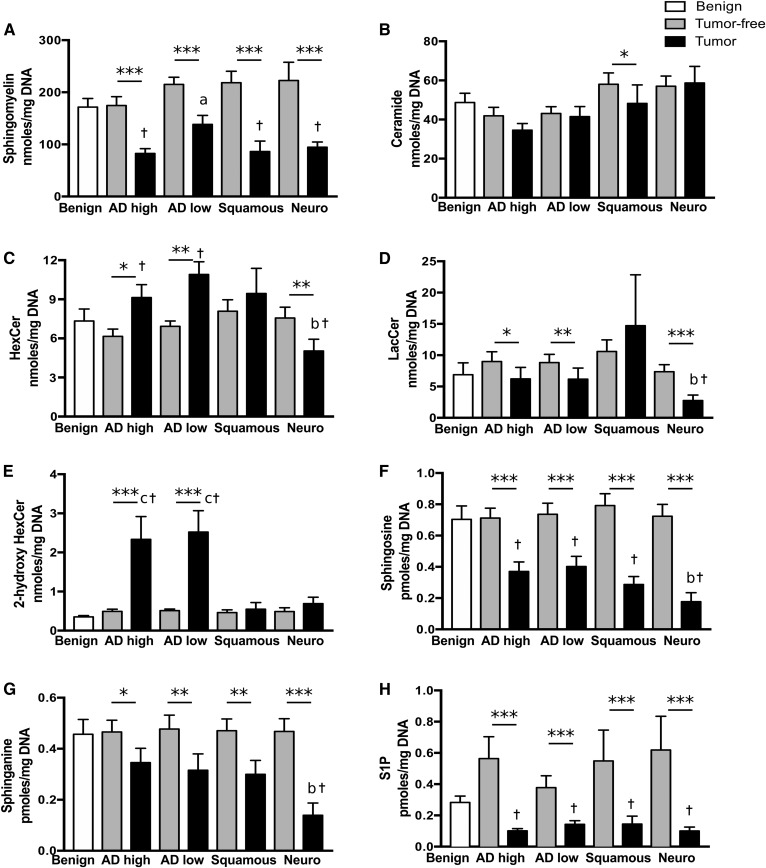
Fig. 1.
Sphingolipids are modulated in different types of lung cancer. Tissue lipids were extracted and sphingolipids were quantified by LC-MS/MS in lung parenchyma of exsmoker patients with benign lung diseases; in tumor and tumor-free lung tissues from patients with high-grade adenocarcinomas (AD high), low-grade adenocarcinomas (AD low), and squamous and neuroendocrine (Neuro) lung carcinomas. The sums of metabolites per lipid class per milligram of DNA are shown for SMs (A), ceramides (B), HexCers (C), LacCers (D); 2-hydroxyHexCers (E), sphingosine (F), sphinganine (G), and S1P (H). Bars represent the average ± SEM, n = 22–25. *P < 0.05, **P < 0.01, ***P < 0.001 tumor compared with tumor-free tissue. aP < 0.05 versus AD high and squamous, bP < 0.05 versus AD high, low, and squamous, cP < 0.05 versus squamous and neuro, †P < 0.05 versus benign.

