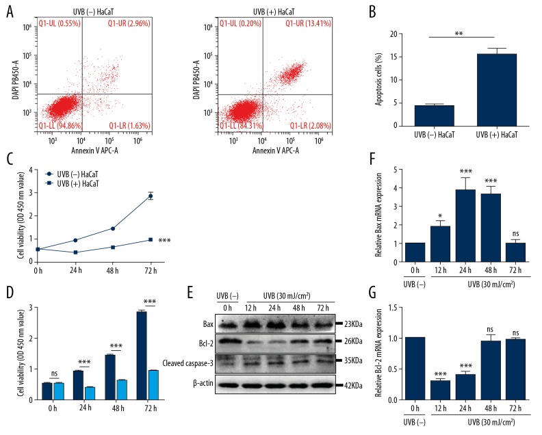
Figure 1.
UVB irradiation inhibited cell proliferation ability and increased apoptosis. (A, B) The proportion of UVB-induced apoptosis was measured by flow cytometry. Bars stand for mean ±SD. ** P<0.01 versus sham-irradiated HaCaT cells [UVB (−) HaCaT], n=3/group. (C, D) At 6, 12, 24, 48, and 72 hours post-UVB (30 mJ/cm2), the cell proliferation ability of UVB-irradiated HaCaT cells was tested using CCK-8 assay. Bars stand for the mean ±SD. *** P<0.001, ns P>0.05 versus UVB (−) HaCaT cells, n=3/group. (E) HaCaT cells were harvested at different time point after UVB irradiation. The protein expression of Bax, Bcl-2, and cleaved caspase-3 were tested using western blotting method; data versus UVB (−) HaCaT cells, n=3/group. (F, G) The relative mRNA expression levels of Bax and Bcl-2 to GAPDH were tested by qRT-PCR at 6, 12, 24, 48, and 72 hours after UVB irradiation. Bars stand for the mean ±SD. *** P<0.001, * P<0.05, ns P>0.05 versus UVB (−) HaCaT cells, n=3/group. SD – standard deviation; UVB – ultraviolet B; CCK-8 – Cell Counting Kit-8; qRT-PCR – quantitative reverse transcription polymerase chain reaction.

