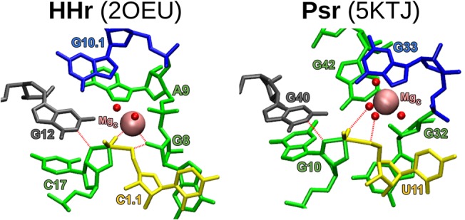
FIGURE 8.

MD models of each crystal equilibrated in a water box as a transition-state-mimic with Mg2+ bound at each ribozyme's C-site. Color-coding corresponds to Figure 7, where the general base guanine is gray. The γ, β2, and δ interactions are labeled with red dashed lines.
