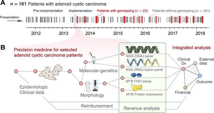
Figure 1.
Clinically integrated diagnostics and analytical workflow. (A): Timeline in days, indicating the diagnosis of adenoid cystic carcinoma (ACC) between 2011 and 2018 by black lines (patients without genotyping) as well as 20 ACC cases, that were sequenced within that timeframe (red lines, patients with genotyping). (B): Analysis of 20 clinically performed formalin‐fixed paraffin‐embedded tissue samples and corresponding clinical data. We integrated five separate diagnostic components including (a) histomorphology, (b) MYB immunohistochemistry, (c) MYB break‐apart FISH, (d) an NGS DNA‐based gene panel, and (e) an RNA‐based NGS panel for fusion detection. For outcome assessment, we employed 201 publicly available (external) data.
Abbreviations: Clinical Dx, clinical diagnostics; FISH, fluorescent in situ hybridization; NGS, next‐generation sequencing.

