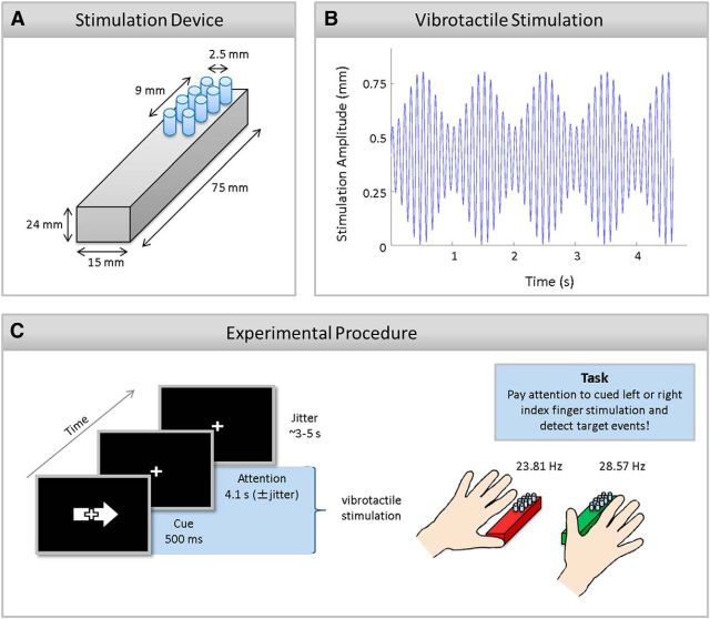
Figure 1.
Schematic of the stimulation device (A), vibrotactile stimulation stream (B), and the experimental procedure (C). A, Lateral top view of the stimulation device for one finger. The stimulation display was composed of eight pins with 2 columns × 4 rows. The diameter of one pin was 1.5 mm and the distance of two neighboring pins 2.5 mm. B, Schematic of tactile stimulation applied to the left and right index finger. A high-frequency carrier signal (142 Hz, for display purposes 10 Hz are plotted) was amplitude-modulated by 23.81 and 28.57 Hz for left and right index finger, respectively (for display purposes 1 Hz is plotted). Note that an event-absent trial is depicted that was predominantly presented and that was used for all performed EEG and fMRI analyses. C, An exemplary trial in which participants were asked to pay attention to the right index finger stimulation. The visual arrow (500 ms) cued participants to allocate and sustain their attention to the left or right index finger stimulation. Participants were asked to detect a target event at the predefined target column on the to-be-attended stimulation. Note that stimulation devices are enlarged in this picture. The pin displays only covered a small part of the distal phalanges. The stimulation devices are depicted in red and green for a better comparison with Figures 2B,C, 4, and 6.

