Figure 2.
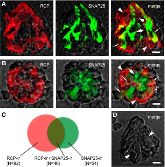
A subset of Presynaptic (Type III) taste cells express RCP. Double immunostaining of isolated and fixed taste buds. Nomarski optics images showing the side (A) and top (B) views of individually isolated taste buds were merged with immunofluorescent confocal micrographs for these micrographs. RCP-immunoreactive (red) taste cells (arrowheads) and SNAP25-immunoreactive (green) Presynaptic (Type III) cells (large arrow) were revealed. Some taste cells exhibit double labeling, suggesting that these Presynaptic (Type III) cells express RCP (small arrows). The optical thicknesses (z-stack) of confocal images are 12 and 21 μm for A and B, respectively. Scale bar, 25 μm. C, Venn diagrams representing the relative proportion of taste bud cells that show RCP (RCP-ir) and/or SNAP25 (SNAP25-ir) immunoreactivity. D, A control taste bud was processed with the omission of the both primary antibodies. Note the absence of immunostaining in cells normally rich with RCP and/or SNAP25 immunoreactivity (arrowheads). Compare with Figure 2A, B. Scale bar, 25 μm.

