
Figure 12.
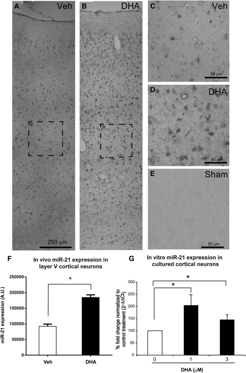
DHA increases miR-21 expression in vivo and in vitro. A, B, Low-magnification images of miR-21 ISH in rats' cerebral cortices ipsilateral to the lesion side 1 d following cervical SCI. C, D, Higher magnification of the dashed boxed in A, B revealed stronger expression of miR-21 in layer V cerebral cortex after DHA treatment. E, Very low miR-21 expression in sham operated animal. F, The quantification revealed that DHA significantly upregulated the level of miR-21 compared with the vehicle group. G, Levels of miR-21 in the cortical neuron culture were determined by qRT-PCR using the TaqMan microRNA assays. The quantitative data were expressed as fold change, in which the RNU6B acted as the endogenous control and the 0 μm DHA as the control for treatment. A significant increase in miR-21 levels with 1 and 3 μm DHA was observed in cortical neuron cultures at 3 DIV. *p < 0.05.