
Figure 7.
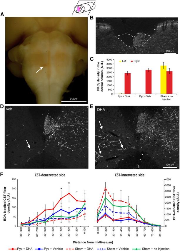
DHA enhances corticospinal axon sprouting following pyramidotomy in mice. A, Ventral view of a mouse brainstem with a unilateral pyramidotomy. Arrow indicates a unilateral lesion of the CST located in the medullary pyramids. B, Lack of PKCγ immunostaining in the left ventral portion of the dorsal funiculus of the cervical spinal cord confirms the unilateral lesion of the CST. C, The intensity of PKCγ immunostaining of the lesioned CST in the cervical spinal cord reveals a significant decrease after pyramidotomy compared with the uninjured CST tract and the sham operated group. D, E, Transverse sections of the cervical spinal cord reveal CST fibers sprouting from the contralateral side across the midline after pyramidotomy in both vehicle and DHA groups. F, Distance-specific analysis of midline crossing fibers shows that the DHA-treated group (red circles) had significantly more midline crossing fibers compared with vehicle (blue squares) and the sham operated group (green triangles) on the CST-denervated side of the cervical spinal cord. Sham operated animals, which received either DHA (red open circles) or vehicle (blue open squares), did not show a significant difference compared with the sham operated group. *p < 0.05, versus the vehicle group. **p < 0.01, DHA versus the vehicle group.