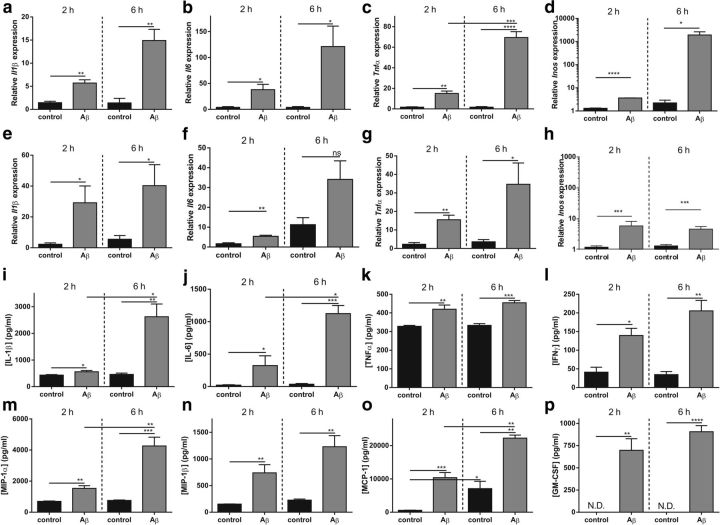
Figure 2.
Cytokine and chemokine analyses of CP, hippocampus, and CSF after intracerebroventricular injection of Aβ1–42 oligomers. a–d, mRNA expression analysis of Il1β (a), Il6 (b), Tnf (c), and Inos (d) in CP after intracerebroventricular injection of Aβ1–42 oligomers (gray) in C57BL/6mice compared with control CP samples (black) (n = 3–4). e–h, mRNA expression analysis of Il1β (e), Il6 (f), Tnf (g), and Inos (h), in the hippocampus after intracerebroventricular injection of Aβ1–42 oligomers (gray) in C57BL/6mice compared with control samples (black) (n = 3–4). i–p, Levels of cytokines (IL-1β, IL-6, TNFα, and IFNγ) and chemokines (MIP-1α, MIP-1β, MCP-1, and GM-CSF) in CSF isolated from C57BL/6 mice injected intracerebroventricularly with scrambled peptide (black) or Aβ1–42 oligomers (gray) (n = 4).

