
Figure 1.
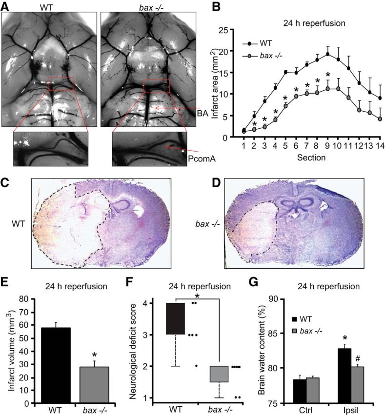
bax deficiency protects against ischemic injury. A, Representative photomicrographs and magnified images of the circle of Willis showing the major blood vessel in WT and bax−/− mice perfused with ice-cold saline followed by cresyl violet solution (A). Arrows: BA, PcomA. There was no significant difference in the mean values of the PcomAs between genotypes (n = 4). B–G, Transient focal ischemia was induced for 60 min in WT (n = 6) and bax−/− (n = 8) mice by a silicon-coated nylon filament that was introduced into the internal carotid artery to occlude the MCA. Surgery was performed in deep isoflurane/N2O anesthesia with controlled ventilation and ischemia and reperfusion were verified by laser-Doppler flowmetry. Mice were killed 24 h after reperfusion and 10 μm coronal sections from each brain samples were collected and taken at 500 μm intervals. Infarct area (B) and infart volume (E) were calculated, as described previously by Gröger et al. (2005). Means ± SEM are shown. *p ≤ 0.05 compared with ischemia-exposed WT controls (Mann–Whitney rank-sum test). The photomicrographs show representative images of cresyl violet/Nissl-stained brain slices from WT and bax−/− mice 24 h after the onset of ischemia. The infarct area remained unstained and is highlighted by dashed lines (C, D). Neurological deficit score (F) was measured 24 h after tMCAo as described in the Materials and Methods session. *p ≤ 0.05 compared with WT controls (Mann–Whitney rank-sum test). Brain water content (G) was quantified and calculated as percentage of contralateral hemisphere to correct for differences in brain size and brain edema. *p ≤ 0.05 compared with contralateral WT controls; #p ≤ 0.05 compared with ipsilateral WT controls (Mann–Whitney rank-sum test).