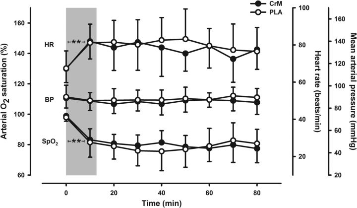
Figure 3.
Autonomic regulation during hypoxia. Arterial oxygen saturation (SpO2) was reduced by 19% during hypoxia. A compensatory 12% increase, on average, in heart rate (HR) occurred, with no changes in blood pressure (BP) detected. Gray shading represents wash-in stabilization period followed by 80 min of hypoxia for CrM (black circles) and PLA (white circles) treatments. Data are mean ± SD; n = 12 (n = 11 for BP). **p < 0.01 for 0 min versus subsequent observations.

