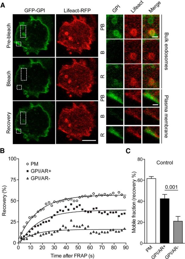
Figure 2.

Actin rings colocalize with GFP-GPI, a plasma membrane probe. A, Chromaffin cells cotransfected with GFP-GPI and Lifeact-RFP were stimulated with nicotine (100 μm) and visualized by time-lapse confocal microscopy. GPI-positive internalized structures were subsequently subjected to FRAP (100% argon laser, 20 iterations), and fluorescence recovery (mobile fraction) was analyzed. Scale bars: A, 10 μm, inset, 1 μm. B, Plasma membrane area were used as a control for recovery. GPI-positive rings that were also positive for Lifeact-RFP actin ring (GPI/Actin Ring Positive: AR+) were shown to recover to a greater extent than GPI-positive rings with no Lifeact-RFP positive actin ring association (GPI/Actin Ring Negative: AR−). C, GPI-positive rings without actin ring association (GPI/AR−) were unable to recover (p < 0.01; n = 16 cells). Error bars are mean ± SEM.
