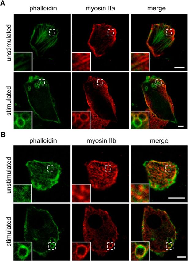
Figure 4.

Myosin IIa and IIb localize to actin rings. A, B, Resting and nicotine-stimulated (100 μm for 8 min) bovine adrenal chromaffin cells were fixed and stained for either myosin IIa (A) or myosin IIb (B) and phalloidin-Alexa Fluor 488. Insets illustrate the presence of myosin IIa and IIb associating with actin at rest (top panels) and in actin ring structures after stimulation (bottom panels). Scale bars, 5 μm.
