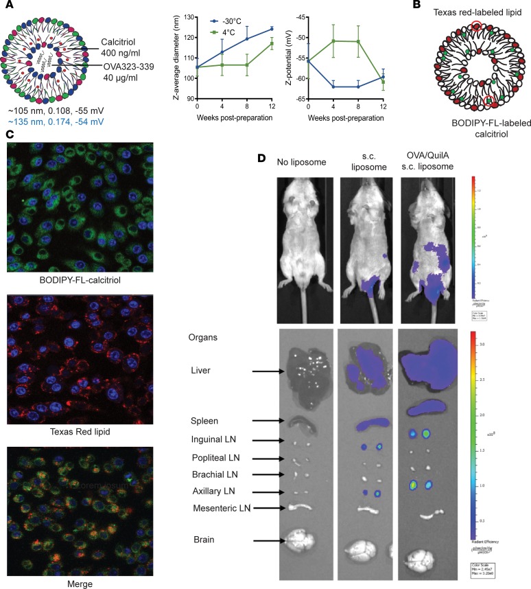
Figure 1. Liposome characteristics and biodistribution.
(A) Size, polydispersity (PDI), and surface charge (black: thin film hydration method, blue: microfluidic method) of liposomes encapsulating calcitriol and OVA323–339 maintained in PBS at –30oC, 4oC, or 25oC for 1 month; n = 3 experiments. (B and C) Single- or dual-labeled liposomes were incubated with RAW 264.7 macrophages and uptake analyzed after 2 hours by confocal microscopy (original magnification, ×60). Blue: DAPI, Green: FL-calcitriol, Red: TXR-lipid. (D) DiR-labeled OVA323–339/calcitriol liposomes were injected s.c. into the tail base of naive or mice primed at tail base with OVA/QuilA 3 days previously. Mice and tissues were analyzed 24 hours after liposome injection by IVIS in vivo imaging. (C and D) Representative of n = 3 per group.

