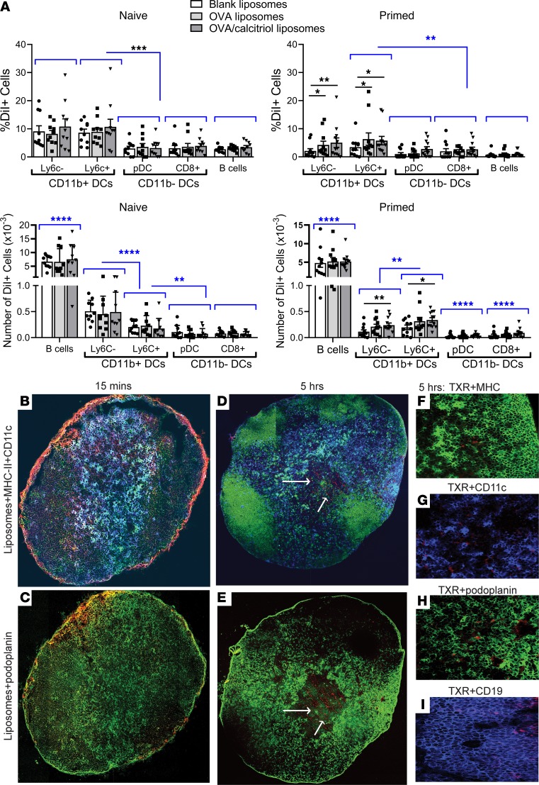
Figure 2. Passive targeting of myeloid DCs in skin dLNs after s.c. injection.
(A) DiI-labeled blank, OVA323–339, or OVA323–339/calcitriol liposomes were injected as in Figure 1D, inguinal lymph node (ILN) cells were stained with CD11c, CD11b, MHC class II, Siglec H, Ly6C, and CD19. DiI+ populations were gated as shown in Supplemental Figure 1A. Total number of DiI+ cells. Flow cytometric data pooled from 10–12 mice/group, representing 2 replicates. *P < 0.05; **P < 0.01; ***P < 0.001; ****P < 0.0001. Analyses shown in blue are for each cell type, uptake of pooled blank, OVA, and OVA/calcitriol liposomes. (B–I) Popliteal lymph nodes from mice primed with OVA/QuilA 3 days before were removed 15 minutes and 5 hours after footpad injection of OVA323–339/calcitriol liposomes incorporating Texas red–DPPC (red) lipid. Lymph nodes were stained with MHC class II–FITC (green) (B, D, and F), CD11c-APC (blue) (B, D, and G), podoplanin-FITC (green, FRC) (C, E, and H), and CD19 (blue) (I). Original magnification, ×20 [B–E, tiled images]; ×20 [F–I, representative of 3 experiments]). Differences in proportion of DiI+ cells compared by mixed-effects analysis with Geisser-Greenhouse correction and Holm-Sidak’s multiple comparison test.

