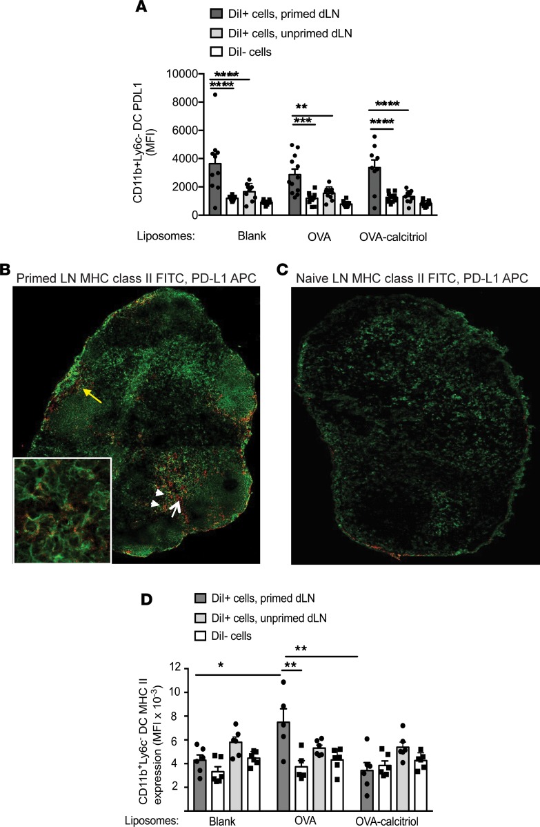
Figure 4. Calcitriol-antigen liposomes, taken up by PD-L1hi CD11b+ primed DCs, suppress DC MHC class II expression.
(A) BALB/c mice were immunized with OVA/QuilA s.c. or were left unprimed and then 3 days later were injected s.c. (both injections at the tail base) with DiI-labeled liposomes. Mean fluorescence intensity (MFI) of PD-L1 in gated DiI+ and DiI– dLN CD11b+Ly6C– DCs 24 hours later. (B) Popliteal lymph node (original magnification, ×10; ×40 [inset]) from a mouse primed with OVA/QuilA 3 days previously was stained with MHC class II–FITC (green) and PD-L1-APC (red) (yellow arrow, afferent lymphatic entry; white arrow, PD-L1+MHC class II– cells; white arrowheads, PD1+MHC class II+ cells.). (C) Popliteal lymph node (original magnification, ×10, relative to isotype control) from a naive mouse stained with MHC class II–FITC (green) and PD-L1-APC (red). Images in B and C are representative of 2 replicates. (D) Mice were treated as in A. MFI of MHC class II in gated DiI+ and DiI– dLN CD11b+Ly6C– DCs 24 hours later. n = 10–12 per group, representative of 2 replicates. *P < 0.05; **P < 0.01; ***P < 0.001; ****P < 0.0001, ANOVA with Sidak’s multiple comparison test.

