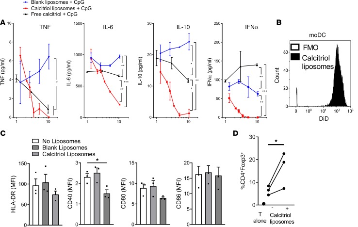
Figure 6. Calcitriol liposomes modulate human DC phenotype and function.
(A) Human PBMCs were stimulated for 18 hours with 10 μg/ml CpG in the absence or presence of varying dilutions of blank liposomes, liposomes encapsulating 400 ng/ml calcitriol and 40 μg/ml OVA323–339, or equivalent final concentrations of free calcitriol. TNF, IL-6, IL-10, and IFN-α concentrations were measured in supernatants. Representative of 2 experiments. *P < 0.05, **P < 0.01, ***P < 0.001, ANOVA at highest calcitriol concentration. The percentage of dead cells was <0.3% for all conditions at highest final calcitriol concentration. (B) Monocyte-derived DCs generated from 3 healthy human PB donors were incubated without or with 0.05% (5 nM calcitriol final concentration) v/v DiD-labeled liposomes for 2 hours and washed, and then DiD fluorescence was analyzed by flow cytometry. (C) Liposome-treated DCs were incubated with 100 ng/ml lipopolysaccharide for 48 hours and then stained with HLA-DR, CD40, CD80, and CD86. *P < 0.05, ANOVA. (D) Liposome-treated DCs were incubated with allogeneic CD4+ T cells (ratio 1:10) for 7 days. The percentage of CD4+Foxp3+ allogeneic T cells identified by flow cytometry is plotted for each donor pair. *P < 0.05, paired t test.

