
Figure 1.
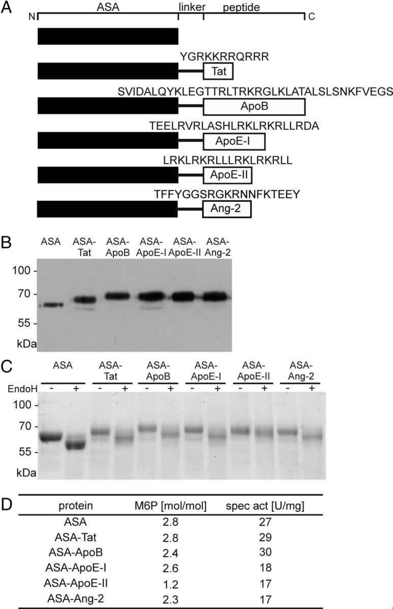
Biochemical characterization of ASA fusion proteins. A, Schemes of ASA fusion proteins. Amino acid sequences of the peptide vectors are given in the one-letter code. Domains are not drawn to scale. B, Western blot analysis. Fifty nanograms of purified ASA and 2.5–10 μl of conditioned medium from CHO-S cells stably expressing ASA fusion proteins were loaded per lane and reacted with a polyclonal anti-human ASA antibody. C, Deglycosylation of ASA fusion proteins with EndoH. Digested and undigested protein was separated by SDS-PAGE and stained with PageBlue Protein Staining solution (Thermo Scientific). D, M6P contents and specific activities as indicated.