
Figure 4.
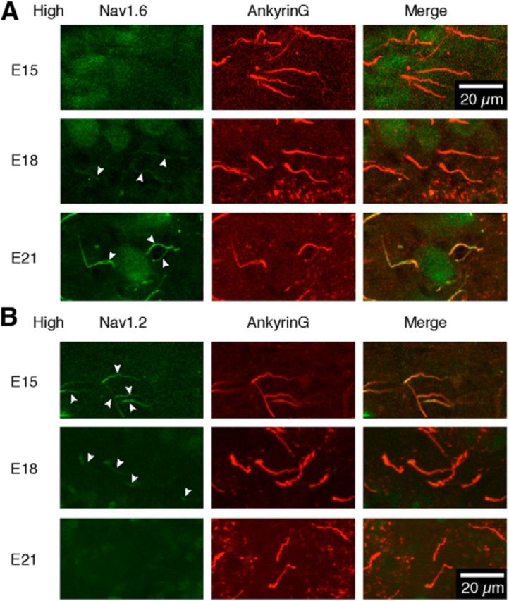
Developmental switching of Nav channel subtypes. A, B, Immunostainings of ankyrinG (red, middle) and either Nav1.2 (A) or Nav1.6 (B) (green, left) were made at the high-CF region and overlaid (right). Note that the AIS was immunopositive for both Nav1.2 and Nav1.6 at E18 (arrowheads).