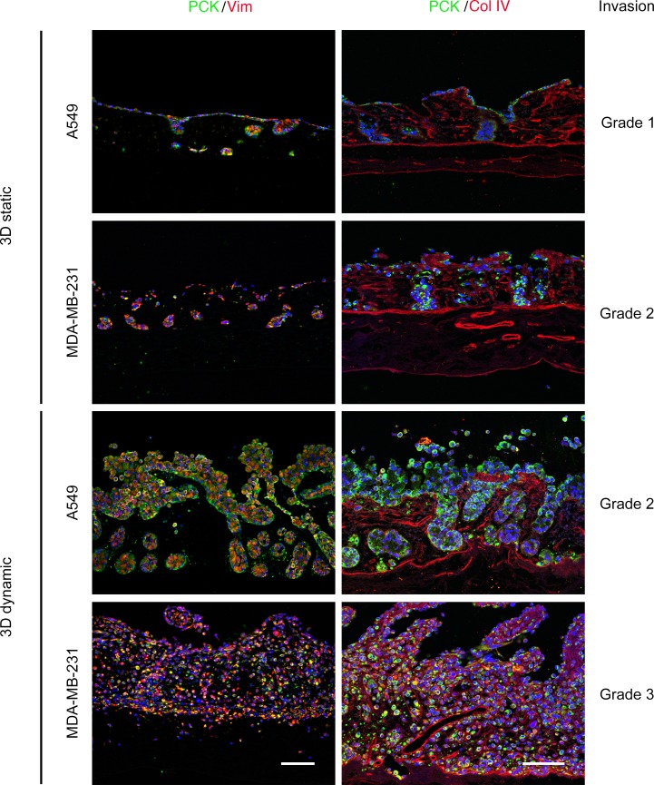
Figure 1. Invasive growth of A549 lung cancer and MDA-MB-231 breast cancer in 3D culture.
The tumor cell lines A549 (lung cancer) and MDA-MB-231 (breast cancer) were cultured on SISmuc scaffolds under static and dynamic culture conditions. Tumor composition and architecture were evaluated by immunofluorescence staining Left column: pan-cytokeratin (PCK, shown in green) and vimentin (Vim, shown in red). Nuclei are counterstained with DAPI (blue). Right column: PCK (green) and collagen IV (Col IV, red). Nuclei are counterstained with DAPI (blue). Scale bars: 100 μm (lower images, representative for all images of a column). Grading was performed according to the scheme presented in Table 1.

