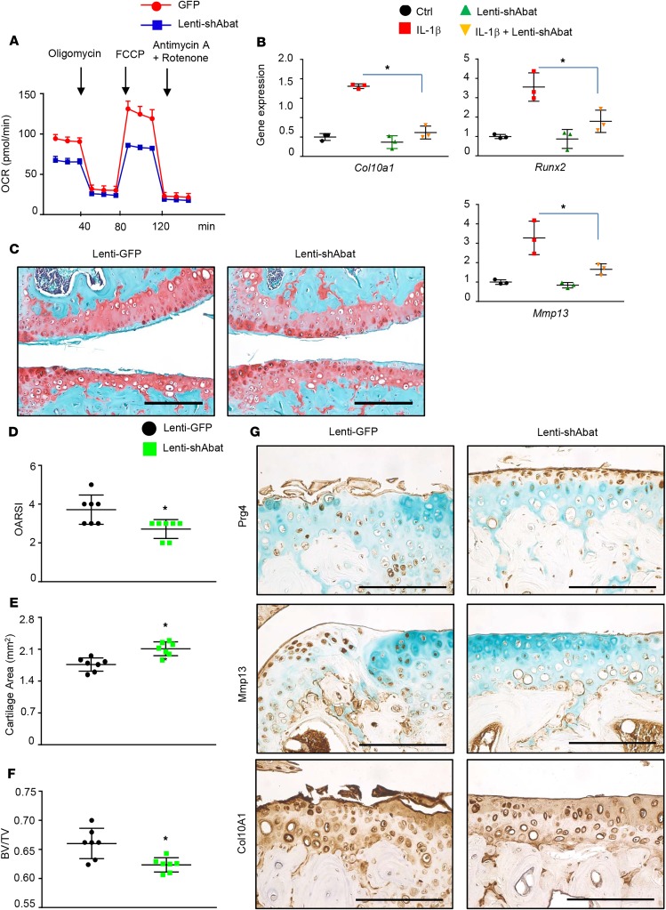
Figure 3. Abat inhibition attenuates chondrocyte hypertrophy and protects cartilage degeneration following surgical induction of OA.
(A) Mitochondrial respiration was measured in primary articular chondrocytes treated with lenti-shAbat (Abat LOF) or lenti-GFP (Ctrl) (n = 8). (B) Real-time qPCR analyses of gene expression from articular chondrocytes transduced with either lenti-shAbat (Abat LOF) or control virus following IL-1β treatment (n = 3). (C) Representative images of histological sections from control or Abat LOF knee joints at 10 weeks following MLI surgery (n = 7). Quantification of histological assessment by OARSI scoring (D) and cartilage area (E) (n = 7). (F) BV/TV ratios of tibial subchondral bone were calculated from the micro-CT images (n = 7). (G) Immunohistochemical analyses for Prg4, Mmp13, and Col10A1 on knee sections of control and Abat LOF cartilage. *P < 0.05 by 2-tailed Student’s t test (A and D–F) and ANOVA with post hoc test (B). Scale bars: 50 μm.

